

Intervertebral disc
The intervertebral disc (IVD) is a highly hydrated, complex, fibrocartilaginous structure critical to the kinematic and biomechanical function of the spinal unit. The central nucleus pulposus (NP), a relatively amorphous, highly hydrated tissue, is encircled by the annulus fibrosus (AF), a complex layered composite of collagen lamellae. Bounding the NP and AF superiorly and inferiorly, the bone-cartilaginous endplates provide the pathway for regulation of nutrient and metabolic byproducts transport from the disc. Under applied load, the AF exhibits anisotropic, non-linear and time-dependent mechanical behavior, differing by orders of magnitude in the tensile stiffness along the lamellar anatomical planes. This behavior and the fine fiber network interconnecting adjacent lamellae are critical to the function of the AF and the IVD's ability to carry functional loading applied to the spine.
With age and degeneration, the NP loses water and proteoglycans; the NP and AF demarcation fades, with the AF showing a decreased number of lamellae, degradation of their structure and changes in the collagen distribution. The resulting loss of NP pressure and altered AF mechanics change the stresses and strains within the IVD that may affect the local failure of the AF, acting as a risk factor for IVD failure and have been postulated to be part of the mechanism underlying disc-affected low back pain.
Clinical MRI is the primary non-invasive diagnostic tool for diagnosing disc pathology, grading the disc degenerative state and identifying compressive lesions. At present, the diagnosis is based on a subjective, qualitative assessment of disc degeneration on T2-weighted MR imaging (Pfirrmann classification: loss of signal due to loss of hydration (NP), loss of anatomical distinction between the NP and AF, herniations, high-intensity zones (NP) and gross changes in shape). However, these signs appear only at later stages and are common in people with no symptoms.
Our group aims to develop quantitative MR and computational methods to interrogate the association between changes in disc structure and composition and its mechanical function from clinical imaging. The work follows three lines:
- MR diffusion as a marker of disc function. Whether diffusion MR detects the spatial structural degradation of the disc tissues as a driver for loss of mechanical performance and whether these metrics identify aging- vs. disease-related disc degeneration better than measures of T2 values.
- Degeneration and spinal mechanics. How disc degeneration changes the response of the spine to high-rate loading, caused by a fall event in frail and cancer patients, and its risk of fracture.
- Image-based computational models. Finite element (FE) models of the disc built from MR geometry, composition and fiber architecture. We aim to use these models to support the development of Machine- and Deep-learning methods for assessing disc function.
Current research
Diffusion tensor imaging of annulus fiber architecture in intact human discs [7]. Degeneration reduces the number of lamellae, disrupts fiber organization and concentrates stress in the inner and posterior annulus, where tears often begin. With the Laboratory of Mathematics in Imaging at Brigham and Women's Hospital, we are investigating the application of high-resolution DTI to map this architecture directly in intact discs.
Early findings:
• In the youngest disc (42 years), DTI clearly separated annulus and nucleus and resolved individual lamellae, with fibers alternating in direction between adjacent layers, as in histology (Figure 1)
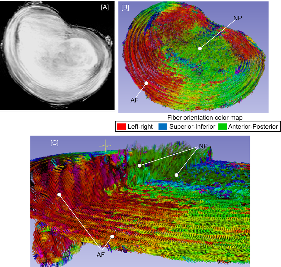
Figure 1: Axial T2 image [A] and corresponding DTI image (1932m*500m), encoded to illustrate the change in fiber orientation as a function of the circumference of the disc [B]. Multi-planar representation [C) shows interlaminar fiber orientation with our of plane fibers in the AF and largely in plane diffusivity in the NP region (B and C).
Comparison of a grade III vs Grade IV (Figure 2) shows that in in older, more degenerated discs (grade IV), DTI showed loss of lamellar definition, disorganized annulus fibers, discontinuities within annulus and nucleus, and blurring of the annulus–nucleus boundary.
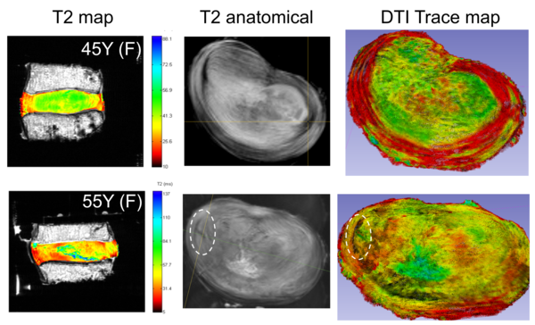
Figure 2.A. Spatial diffusivity shows degeneration affecting loss of lamellar definition, increased heterogeneity of diffusivity within the AF and NP and loss of AF and NP tissue | 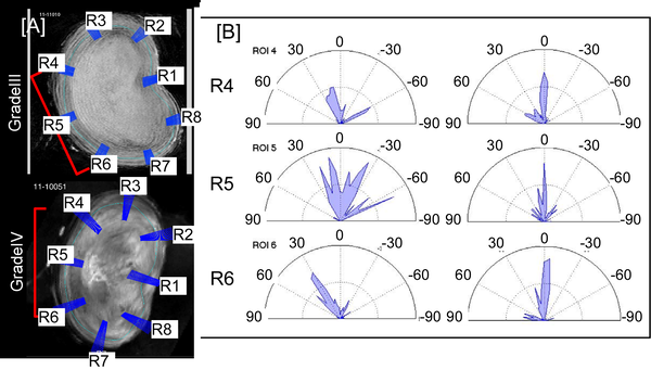
Figure 2.B Computation of fiber angle as a function of IVD circumference shows higher degeneration causing loss of anatomical lamellar fiber orientation, suggesting higher structural damage within the lamellae. |
|---|
The ability to noninvasively obtain precise, quantitative information on the derangement of the AF architecture may improve the fundamental understanding of the effects of degeneration on the structure-function relationship within the disc and allow the development of diagnostic and predictive methods for clinical evaluation of disc disorders associated with injury, degeneration, and aging. Our current work focuses on:
- Developing protocols for imaging under controlled force and /or displacmentimaging within the MR scanner
- Transitioning and optimizing our DTI protocol for IVD imaging in a clinical scanner to allow both research and transition to patient studies.
- Converting the tensor data into anatomically detailed FE models to study how degeneration changes the way the disc fails under functional loading.
Previous Research
A. MR Diffusion and aging [1].
T2-weighted MRI reflects mainly the disc's water and proteoglycan content, and its links to disc composition and mechanics are inconsistent. Diffusion MR measures how freely water moves through the tissue, and so probes its microstructure. We asked whether diffusion tracks disc function and aging more closely than current quantitative T2 protocols.
Study: Ten thoracic and lumbar disc joints from donors aged 37–81 years were imaged at 9.4 T with T2 and apparent diffusion coefficient (ADC) protocols. The nucleus and annulus were segmented, and the mean and variation (COV) of each measure were tested against age.
Key findings:
- With age, mean ADC fell (P < 0.001) and its variation within the disc rose (P < 0.001), in both nucleus and annulus.
- ADC was a more sensitive marker of age-related change than T2.
- Lumbar discs differed from thoracic discs in both ADC and T2, suggesting that the mechanical environment shapes how disc tissues age.
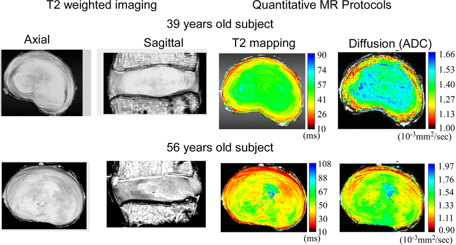
Figure 3. Axial and sagittal images with T2 and ADC maps for a 39-year (grade II) and a 56-year (grade III) lumbar disc, showing age-related change in both nucleus and annulus [1].
B. MR diffusion interrogates disc mechanics [1, 2]
Cadaveric human lumbar discs were imaged at 4.7 T at 200 N (representing baseline) and 800 N (representing free-standing) compressive loads. The disc’s dynamic and viscoelastic properties were measured prior to imaging in cyclic and creep tests, and regression models related the change in T2 and diffusion to disc mechanics.
Key findings:
- Compression reduced the disc’s mean diffusivity from 11.3 to 9.7 × 10⁻⁴ mm²/s (P < 0.001), with little change in T2.
- Diffusivity correlated with the disc’s viscoelastic damping (r² = 0.45) and instantaneous stiffness (r² = 0.44); T2 did not.
- Diffusivity in the nucleus was higher than in the annulus, and T2 and diffusivity agreed at low load but not at high load.
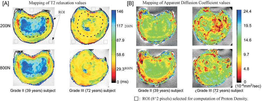
Figure 4. Spatial maps of T2 (A) and apparent diffusion coefficient (B) for a grade II (39-year) and a grade III (72-year) disc at 200 N and 800 N compression. Loading reduces diffusivity across the disc [1].
C. Image-based finite element models of the disc [3, 4]
Current FE models of the disc assume the shape, composition and fiber layout of a healthy disc, which limits their use for studying degeneration. In collaboration with the University of Bern (Prof. Philippe Zysset's group), we investigated whether directly implementing disc-specific, spatial fiber information, derived from our DTI MR protocols, improves the FE models' prediction of the spinal unit multi-planar mechanical response.
MRI-based geometry, composition and fiber architecture [3].
An L2–L3 disc joint was imaged at 9.4 T with proton density and DTI sequences. The proton density image defined the disc's shape and a smooth nucleus–annulus transition. From the DTI data, we extracted the main diffusion direction at each point to set the local collagen spatial fiber angle and the information used to derive a continuum mechanics-based formulation for the NP and AF. The spinal unit mechanical behavior was measured using a custom, computerized, active, 6-DOF test system to evaluate the spinal unit response in tension, compression, flexion, extension, lateral bending and axial rotation.
Key findings:
- DTI revealed the layered structure of the annulus, with fiber angles of about 25–50° to the transverse plane that became less organized toward the nucleus.
- The MRI-based FE model reproduced all six measured load–displacement curves, a first proof of concept for patient-specific disc models.
- Incorporating disc-specific spatial AF information showed clear improvement in prediction of the spinal unit multi-planar mechanical response compared to the current computational model assumption of a fixed, literature-derived "global" value.
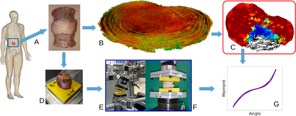
Figure 5. Study overview. A human L2–L3 disc (A) was imaged with proton density and DTI at 9.4 T (B) to build a disc-specific FE model (C). The disc was tested in six loading modes (D–F), and the model was calibrated against the measured curves (G) [3].
D. Disc degeneration and spinal mechanics [5, 6]
In older adults, 30–50% of vertebral fractures follow a fall. The peak force reaching the vertebra in a fall depends on the stiffness and damping of the spine, which the disc largely sets. Yet fracture risk is judged from bone density alone, measured under slow, quasi-static assumptions. We studied how disc degeneration changes the response of the spine to high-rate loading and its failure strength.
Association of MR-derived (T2) disc degradation with the IVD impact response [5].
This project investigated the effect of disc degeneration, evaluated via T2-weighted MR data, on the functional spinal unit stiffness and damping behavior. Nineteen L1–L3 human spine segments were struck with a custom pendulum (20.9 kg end mass) under increasing preload. A Kelvin–Voigt model fitted to the post-impact oscillations gave each segment's stiffness and damping.
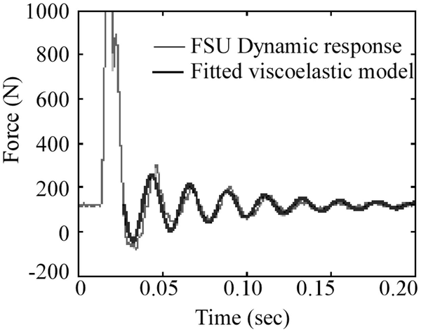
Figure 6. Force response of a spine segment to impact, with the fitted Kelvin–Voigt viscoelastic model used to compute stiffness and damping [5].
Key findings:
- Spine segments showed an under-damped, second-order response, with stiffness of 17.9–754.5 kN/m and damping of 133.6–905.3 Ns/m.
- Stiffness, but not damping, rose sharply with preload (P < 0.001).
- Disc degeneration, seen as lower T2, was weakly linked to stiffness at low preload.
This project highlighted that dynamic models of falls and vibration need to account for non-linear stiffness.
Disc hydration is a strong predictor of spine failure under fall-like loading [6].
Seventeen L1–L3 segments were imaged with MRI and DXA, impact-tested, and loaded to failure at a high rate simulating a fall. Disc hydration and geometry were measured from proton density and T2 images.
Key findings:
- Higher bone density and higher MR-estimated disc hydration both predicted greater ultimate strength (P < 0.01 for both).
- Higher damping was linked to lower strength (P < 0.05); T2 values were not linked to strength.
- Adding disc hydration to bone density strengthened the prediction of failure load by 20% (P < 0.001).
These studies show that the disc's state is an independent predictor of spinal failure under fall conditions and should be part of fracture-risk assessment in the aging spine.
Publications
- Alkalay RN, Hackney DB. Diffusion based MR measurements correlates with age-related changes in human intervertebral disks. Clin Biomech 2019;61:38–45. doi:10.1016/j.clinbiomech.2018.06.007
- Alkalay RN, Burstein D, Westin C-F, Meier D, Hackney DB. MR diffusion is sensitive to mechanical loading in human intervertebral disks ex vivo. J Magn Reson Imaging 2015;41:654–664. doi:10.1002/jmri.24624
- Stadelmann MA, Maquer G, Voumard B, Grant A, Hackney DB, Vermathen P, Alkalay RN, Zysset PK. Integrating MRI-based geometry, composition and fiber architecture in a finite element model of the human intervertebral disc. J Mech Behav Biomed Mater 2018;85:37–42. doi:10.1016/j.jmbbm.2018.05.005
- Stadelmann MA, Stocker R, Maquer G, Hoppe S, Vermathen P, Alkalay RN, Zysset PK. Finite element models can reproduce the effect of nucleotomy on the multi-axial compliance of human intervertebral discs. Comput Methods Biomech Biomed Engin 2020;23(13):934–944. doi:10.1080/10255842.2020.1773808
- Wilson SE, Alkalay RN, Myers E. Effect of the degenerative state of the intervertebral disk on the impact characteristics of human spine segments. Front Bioeng Biotechnol 2013;1:16. doi:10.3389/fbioe.2013.00016
- Alkalay RN, Vader D, Hackney DB. The degenerative state of the intervertebral disk independently predicts the failure of human lumbar spine to high rate loading: an experimental study. Clin Biomech 2015;30(2):211–218. doi:10.1016/j.clinbiomech.2014.09.016

