

Musculoskeletal Modeling

Vertebral fractures are likely to occur if a daily task-related load exceeds the vertebra's capacity to carry such loading, i.e exceeds its structural strength. Most of those loads originate from the trunk muscles, which hold the body upright and balance the trunk for any loading held in the hands. The magnitude of such loads depends on body size, spinal curvature, vertebral level, and the task performed. Measuring muscle-generated forces and resulting vertebral loading faces significant technical difficulties and requires a unique laboratory setting, both impractical when evaluating infirm cohorts of cancer patients with spinal metastasis. 

Derived from clinical imaging, spine musculoskeletal models incorporate personalized anatomy (location and cross-sectional areas) of the flexor, extensor and stabilizing thoracic and abdominal muscles and the anatomy and strength of the vertebral column and can be used to evaluate the loading carried by each vertebra under a daily task. This data is used to derive the level's load-to-strength ratio (LSR), a metric for the patient's risk of fracture.

In patients with metastatic spine disease, tumors change vertebral strength, with the systemic effects of cancer affecting sarcopenia and cachexia, both hallmarks of cancer. Yet, clinical prediction tools used to judge patients' risk of spinal instability and, indirectly, risk of vertebral failure, such as the Spinal Instability Neoplastic Score (SINS), base their prediction on how a lesion appears on imaging but do not consider either the lesion's vertebral mechanics or the loading applied to this vertebra during daily life. 

Our group, working with Dennis Anderson, uses individualized musculoskeletal models of the spine to estimate the loading and resulting LSR in cancer patients during everyday tasks as a metric of their vertebral fracture. We first applied this approach to cadaveric spines from donors with cancer [1] and then to routine clinical CT from 135 patients planned for radiotherapy at Dana-Farber Cancer Institute (DFCI) and Brigham and Women's Hospital (BWH) [2]. In both studies, the cancer spines were compared with a population-based reference group from the Framingham Heart Study. The models draw on our finite element work for vertebral strength and on our image analysis and AI tools
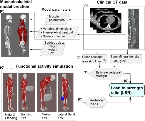
Musculoskeletal-modeling-methodology-work-flow

Figure 1. Workflow for establishing musculoskeletal spine models for personalized assessment of cancer patients' estimated task- and vertebral-specific loading. The patient/subject CT data is used to extract the subject-specific information (A: sex, height/weight, muscle morphology, and spinal curvature). This information is used to adjust and personalize our generic spine models (B). Simulating different postures and loading,   we apply static optimization to predict muscle forces and calculate spinal joint loading using the joint analysis tool in OpenSim ™. The outcome of this analysis is individual vertebral loading (D). Combined with CT estimation of vertebral strength (E - F), we compute vertebral-specific load-to-strength ratio (LSR) (G).
Previous studies

Load-to-strength ratios in cadaveric metastatic spines [1]

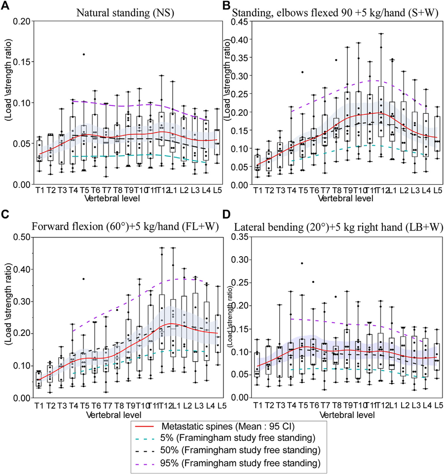
Using our musculoskeletal modeling in 11 cadaveric spines from donors with metastatic spine cancer and in a non-cancer cohort (250 participants) from the Framingham Heart Study. We found osteosclerotic and mixed vertebrae with lower LSRs than sex-matched and age-similar normative values (FHS), results largely driven by the vertebrae's higher strength. By comparison,  osteolytic vertebrae had higher LSRs, up to 107% above normative values in the lumbar spine. Unexpectedly, we found vertebrae with no lesion visible on CT also had higher LSRs than normal, suggesting that fracture risk may be raised even at levels that appear healthy.

Effect of simulated daily tasks on the change in vertebral compressive.

Figure 2. Effect of simulated daily tasks on the change in cancer patients vertebral load-strength ratio (LSR) for representative daily tasks. (A) Nautral standing, (B) standing + holding a weight (elbows flexed 90° with 5 kg in each hand), (C) 40° trunk flexion + 5 kg in each hand, and (D) 20° trunk lateral bending to the right + 5 kg in the right hand.  The dotted lines represent the 5th percentile (green), median, 50%, (black), and 95th percentile (purple) of the computed LSR values for each activity modeled using the data obtained for the FHS cohort (Mokhtarzadeh, et al,.,  (2021). J. Bone Min. Res. 36 (4), 704–711. doi:10.1002/jbmr.4222.

Load-to-strength ratios in patients planned for radiotherapy [2]

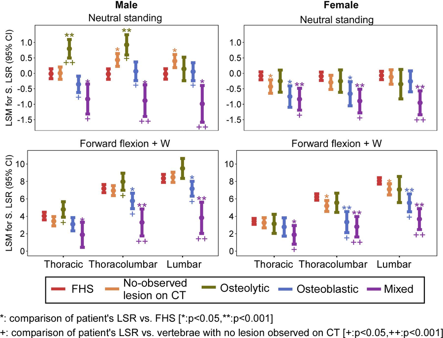
We extended the approach to a study of 135 patients treated at Dana-Farber Cancer Institute and Brigham and Women's Hospital. For this study, we computed LSRs for 1,508 vertebrae and compared computed LSR with those computed for 246 Framingham participants [2]. We found that lesion quality affects the patients' LSRs. Vertebrae with mixed lesions had lower LSRs than controls in both sexes, while osteolytic vertebrae had higher LSRs in men but not in women. Uniquely, we found the difference was driven partly by the effect of primary cancer on the estimated muscle-generated forces and by body weight. Of importance was the finding that Vertebrae with no visible lesion differed from FHS age-similar and sex-matched participants, with the difference in opposite directions in men and women. These findings indicate that cancer may affect the biomechanics of the whole spine, with levels considered "normal" likely at increased risk of failure depending on the task performed. 
 

Effect of simulated daily tasks on the change in vertebral LSR-patient study.

Figure 3. Comparison of least squares mean (LSM) differences in standardized spine LSRs between spine metastases patients and Framingham Heart Study (FHS) participants, presented for men and women and grouped by lesion type and region [2]. Lesion type significantly affected LSRs per region and task, with marked differences observed for these effects between men and women as compared to FHS participants.

Current directions

  • Our group is working to evaluate whether LSRs predict pathologic fracture in patients followed after radiotherapy, and to define risk thresholds by spinal region and lesion type.
  • We are working to add deep learning-based automated trunk-muscle segmentation, CT [3] and MRI, to increase the automation and accuracy of patient-specific models and extend our model to true 3D modeling.
  • We are working to establish our models as part of 3Dslicer to allow open-source access for evaluating patients.
  • We aim to implement more advanced image-analysis assessment tools for muscle quality to refine and extend our model muscle force estimation in cancer and elderly patients. As part of this development, we aim to introduce a registration method to evaluate the temporal effect of cancer treatment (radiotherapy) on muscle properties and test its effect on cancer patients' frailty, spinal stability and fracture risk.

Publications

  1. Anderson DE, Groff MW, Flood TF, Allaire BT, Davis RB, Stadelmann MA, Zysset PK, Alkalay RN. Evaluation of load-to-strength ratios in metastatic vertebrae and comparison with age- and sex-matched healthy individuals. Front Bioeng Biotechnol. 2022;10:866970. doi:10.3389/fbioe.2022.866970
  2. Anderson DE, Keko M, James J, Allaire BT, Kozono D, Doyle PF, Kang H, Caplan S, Balboni T, Spektor A, Huynh MA, Bouxsein ML, Hackney DB, Alkalay RN. Metastatic spine disease alters vertebral load-to-strength ratios in cancer patients compared to healthy individuals. JOR Spine. 2026;9(1):e70111. doi:10.1002/jsp2.70111
  3. Hong V, Pieper S, James J, Anderson DE, Pinter C, Chang YS, Bulent A, Kozono D, Doyle P, Caplan S, Kang H, Balboni T, Spektor A, Keko M, Kikinis R, Hackney DB, Alkalay RN. Automated segmentation of trunk musculature with a deep CNN trained from sparse annotations in radiation therapy patients with metastatic spine disease: an observational study. Front Bioeng Biotechnol. 2026;13:1707724. doi:10.3389/fbioe.2025.1707724