

Vertebral Augmentation
Vertebral Augmentation-Current research
Percutaneous cement augmentation (vertebroplasty, Kyphoplasty) is widely used minimally invasive medical procedure to relieve pain and stabilize vertebrae fractured by osteoporosis or weakened by lytic metastases. Clinical studies report rapid pain relief in many patients, yet how much mechanical competence the procedure actually restores to a failed vertebra, and whether that restoration holds under everyday loads, remains poorly understood. Specifically, how the the lesion mass parameters and its interaction with the bone environment and/or existence of fractures or vertebral deformity, determines the augmentation procedure outcomes, remains similarly unclear. Our group aims to address this gap by combining experimental, imaging and computational approaches for quantifying the multi-planar structural response of failed human vertebrae before and after augmentation, under combined compression and forward-bending loads that reproduce the clinical anterior-wedge fracture.
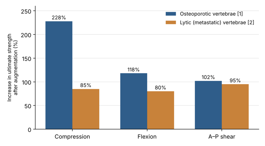
Figure 1. Augmentation strengthens failed osteoporotic vertebrae more than failed osteolytic vertebrae. Percent increase in ultimate strength after augmentation, relative to the failed state, as reported in [1] and [2].
Research program:
- Augmentation of failed osteoporotic vertebrae.
- Augmentation of failed metastatic (osteolytic and mixed lesion) vertebrae.
- Computational modeling of the augmentation process in metastatic vertebrae.
Current research
Finite element simulation of augmentation in metastatic vertebrae. Our experiments showed that the benefit of augmentation in lytic vertebrae depends on cement volume and distribution, and is limited by extravasation.
We are developing finite element models that simulate the augmentation process in metastatic vertebrae, with the aim of predicting how cement placement and volume affect the structural response of the treated vertebra. These models are a step toward image-based preoperative tools for planning augmentation in patients with metastatic spine disease.
Vertebral Augmentation - Previous research
A.1: Augmentation of failed osteoporotic vertebrae [1]
Vertebral fragility fractures affect up to 500,000 people in the United States each year. Patients are typically treated some time after the injury, once the fractured vertebra has partially recovered. To reproduce this clinical scenario, we developed an anterior-wedge, post-failure recovery model. Nineteen osteopenic thoracolumbar vertebrae (T6–L2) were tested to failure under combined compression and flexion, allowed to recover, re-tested to failure, augmented with PMMA cement through a bipedicular approach, and tested to failure a third time. A six-degree-of-freedom load cell captured the full force and moment response of each vertebra at every stage.
Key findings:
- Augmentation increased compressive strength by 228% and flexion strength by 118% relative to the failed vertebrae, exceeding intact values.
- Augmentation was far less effective in restoring the vertebra's resistance to bending moments (flexion and lateral bending) than to compression and shear.
- The augmented vertebrae showed a distinctly bilinear load response, suggesting that the cement acts as a rigid core about which the remaining damaged bone deforms.
- Augmentation partially restored vertebral body height, but most of this restoration was lost once the vertebra was reloaded.
- The degree of pre-existing wedge deformity predicted height restoration, but not the gain in strength or stiffness.
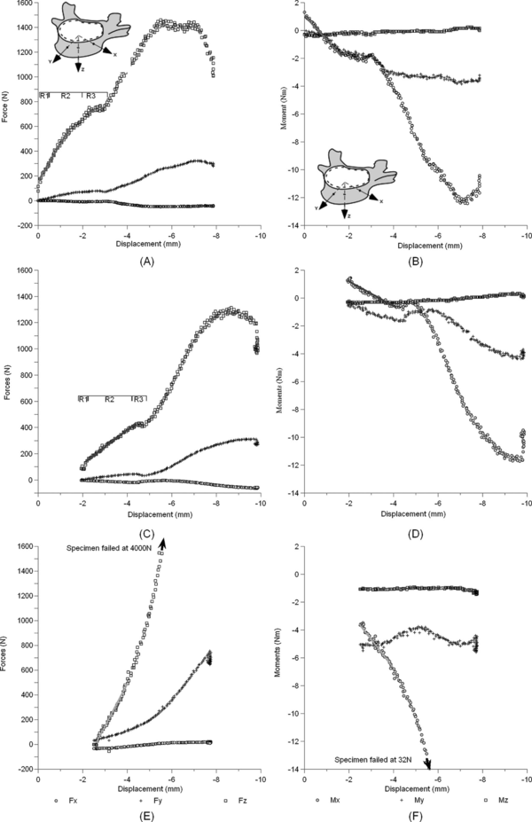
Figure A.1. Force (left) and moment (right) versus displacement for a T11 vertebra: intact (A, B), recovered after failure (C, D) and augmented (E, F). Failed vertebrae show loss of compressive load response and ability to resist forward and lateral bending moments, suggesting a degraded structural response. Post-augmentation the compressive response rises steeply, while bending is restored far less and importantly the pattern of response is significantly modified, suggesting an alteration in the structural response of the treated spinal unit which may expose it to accelerated failure [1].
Our finding fuhrer questioned the procedure efficacy for restoring vertebral body heights, one of the main clinical claims for this procedure
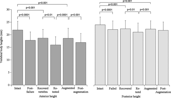
Figure A.2. Anterior and posterior vertebral body heights across test stages. Augmentation restores height, but much of the gain is lost under reloading [1].
Augmentation of failed metastatic vertebrae with lytic defects
B. Augmentation of failed metastatic vertebrae with osteolytic defects
The vertebra is the most common site of skeletal metastasis, and osteolytic lesions markedly reduce vertebral strength. Although augmentation is routinely used for pathologic fractures, little was known about how well it restores the strength of a vertebra that has already failed around the osteolytic lesion. In a prior study, we created a critical, uncontained lytic defect (40% of the vertebral body volume, breaching the cortex) in eighteen osteoporotic lumbar vertebrae (L1–L5) and characterized their failure under compression and forward bending [3]. The failed vertebrae were then augmented with PMMA through both pedicles, filling the defect site first, and re-tested to failure using the same wedge-fracture model [2].
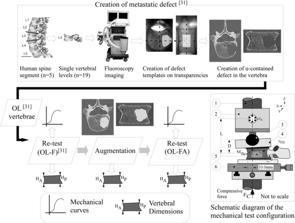
Figure B.1 Experimental workflow. A simulated uncontained lytic defect was created under fluoroscopic guidance; the vertebrae were failed, augmented with PMMA, and re-tested under combined compression and flexion. Vertebral heights and mechanical curves were recorded at each stage [2].
Key findings:
- Augmentation increased the failed osteolytic vertebrae's compressive strength by 85%, flexion strength by 80%, and anterior–posterior shear strength by 95%, with stiffness gains of 40–53%.
- Augmented osteolytic vertebrae reached 50–90% of the strength reported for osteoporotic vertebrae without osteolytic defects, and remained up to 20% weaker than untreated, otherwise normal osteoporotic vertebrae.
- Cement volume correlated with compressive and shear strength, but its benefit was self-limiting: cement extravasated through the defect or fractured endplates in every specimen (mean volume 5.0 cc).
- As in osteoporotic vertebrae, augmentation was less effective in restoring moment-carrying capacity, and height restoration was largely lost under reloading.
- Neither bone mineral density nor vertebral deformity predicted the volume of cement that could be injected.
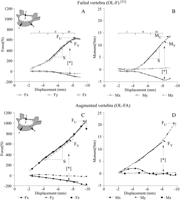
Figure B.2 Force and moment versus displacement for a failed lytic vertebra (A, B) and the same vertebra after augmentation (C, D). Augmentation converts the nonlinear response of the failed vertebra to a near-linear response with no evidence of structural failure [2].
Publications
- Alkalay RN, von Stechow D, Torres K, Hassan S, Sommerich R, Zurakowski D. The effect of cement augmentation on the geometry and structural response of recovered osteopenic vertebrae: an anterior-wedge fracture model. Spine 2008;33(15):1627–1636.
- Alkalay RN, von Stechow D, Hackney DB. Augmentation of failed human vertebrae with critical un-contained lytic defect restores their structural competence under functional loading: an experimental study. Clin Biomech 2015;30:608–616. doi:10.1016/j.clinbiomech.2015.03.024
- Alkalay RN. Effect of the metastatic defect on the structural response and failure process of human vertebrae: an experimental study. Clin Biomech 2015;30(2):121–128. doi:10.1016/j.clinbiomech.2014.10.001

