Routine EM Services
- Consultation
- Tissue Preparation
- Tissue Processing
- Thick/Thin Sectioning
- Contrast Staining
- Service or Independent use of the electron microscope.
- Image Analysis
Getting Started
Because EM projects can be extensive and expensive, the EM Core Facility invites all researchers to meet with core staff before a project is started to discuss the scope of the project, how to procure and fix tissues, timeline, budgetary concerns, and any other general issues which might arise.
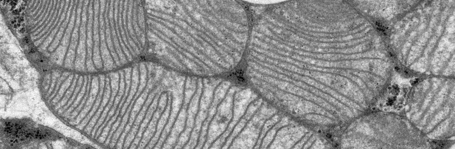
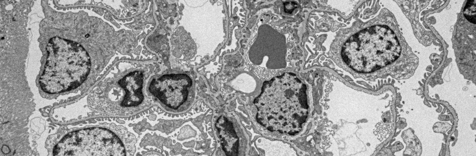
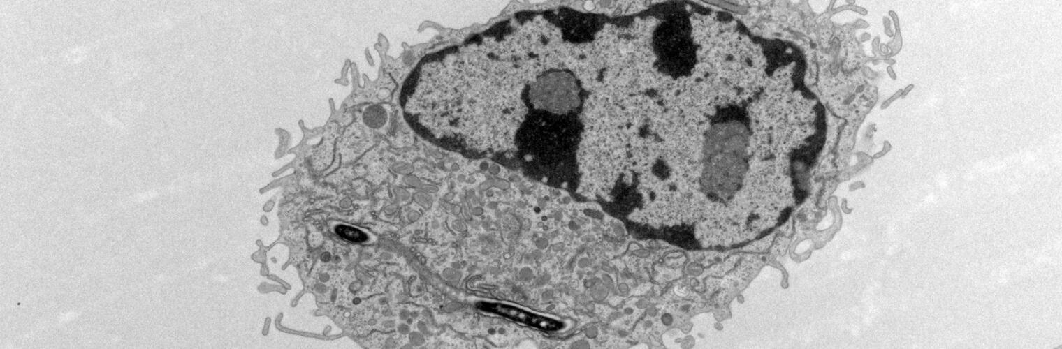
Techology & Equipment
- JEOL 1400
- Installed January 2010 and is state-of-the-art.
- 120kV
- High contrast
- Gatan Orius 11 MegaPixel CCD Cameras for photodocumentation
- SerialEM, IMOD, and Chimera EM Tomography software.