
 On the Cover

Small • November 26, 2018
In article Picoanalysis of Drugs in Biofluids with Quantitative Label‐Free Surface‐Enhanced Raman Spectroscopy, Lev T. Perelman and co‐workers report a quantitative label‐free surface‐enhanced Raman spectroscopy (SERS) method for identifying and quantifying drugs at ultralow concentrations. Self‐assembled gold nanoclusters entrap drug molecules and laser light excites the surface plasmon resonance, yielding a large SERS signal from the molecule trapped near the hot spot, allowing measurements of drugs in urine samples at clinically relevant concentrations...show more

 The Latest Cancer Research News to Cheer About

PARADE • By Catherine Winters • June 16, 2017
Pancreatic Cancer
About one-fifth of pancreatic cancers evolve from cysts, but there’s no easy way to determine if a cyst is benign, pre-cancerous or malignant. Even if a doctor suspects cancer, a biopsy detects only about half of pancreatic cancers. Researchers at Beth Israel Deaconess Medical Center in Boston have developed a tool called light scattering spectroscopy that bounces light off the cyst, analyzing it for structural changes that may signal cancer. While more testing is needed, the technology “could potentially transform the diagnosis of pancreatic cancer by enabling doctors to confirm a malignancy quickly, early and noninvasively and follow cysts that could potentially become cancerous,” says Lev T. Perelman, Ph.D., director of the Center for Advanced Biomedical Imaging and Photonics at Beth Israel...show more
 Light-scattering tool peers into pancreas to find cancer
Discovery • June 21, 2017
NSF-funded basic research leads to technique that could identify potentially cancerous cells with greater accuracy
Pancreatic cancer is difficult to detect early because the pancreas is deep inside the abdomen, making potentially cancerous cells hard to reach and identify without surgery. Researchers funded by the National Science Foundation (NSF) developed a new light-based technique that can identify precancerous and cancerous cysts -- small, fluid-filled cavities in the body -- by piggybacking on a standard diagnostic procedure. "This approach can be called a virtual biopsy, as it does not collect any tissue," said Lev Perelman, professor at Harvard University and director of the Center for Advanced Biomedical Imaging and Photonics ...show more

 NIBIB-funded researchers develop optical biopsy tool to identify early pancreatic cancer

Science Highlights • May 15, 2017
Endoscopic procedure achieves 95 percent accuracy in detection of benign and potentially cancerous cysts
Researchers funded by the National Institute of Biomedical Imaging and Bioengineering (NIBIB) have developed a new tool for detecting early pancreatic cancer. The tool bounces light off targeted tissue to detect structural changes in the tissue, a method called light-scattering spectroscopy (LSS). The researchers performed a series of pilot studies using LSS and accurately distinguished benign cysts, cancerous cysts, and those with malignancy potential 95 percent of the time. The Harvard University team reported their results in the March 13, 2017, issue of Nature Biomedical Engineering...show more
 Brighter cancer probes

Nature Biomedical Engineering Editorial
The early detection of cancer demands translatable light-emitting or light-collecting probes with unprecedented levels of sensitivity and specificity.
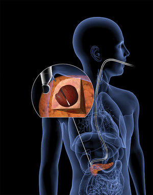
The best clinically available method for the identification of malignant pancreatic cysts is endoscopic ultrasound-guided fine-needle aspiration, yet its specificity is only ∼50%. Lev Perelman and colleagues (article no. 0040) describe a fibre-optic probe, compatible with routine endoscopic-aspiration procedures (see image), that distinguishes weakly scattered photons from cancer tissue over the more broadly diffuse photons from healthy tissue ... show more

 Cancer diagnostics: Light scattering by pancreatic cysts

Nature Biomedical Engineering News and Views
Pancreatic cysts can be detected via the optical-scatter patterns of these submicroscopic sack-like pockets of tissue... show more

 Spectroscopic technique identifies precancerous cysts

IOP Publishing MedicalPhysicsWeb
Light scattering spectroscopy (LSS) performed during an endoscopic ultrasound-guided fine-needle aspiration (EUS-FNA) can accurately identify pancreatic cystic lesions that are potentially malignant. This capability can significantly improve distinction between precancerous and benign cysts and help determine whether surgical resection is needed...show more

 Microscopy shines at BiOS Hot Topics

SPIE Photonics West. Kathy Kincade
Advances in microscopy dominated the BiOS Hot Topics presentations at Photonics West, detailing advances in new techniques that could dramatically influence molecular research, drug development, and clinical diagnostics.
In his talk on "Biomedical Imaging and Spectroscopy with Scattered Light," Lev Perelman of Harvard University Beth Israel Deaconess Medical Center shared his group's research involving CLASS (confocal light absorption and scattering spectroscopic microscopy). This unique combination of confocal microscopy and light-scattering spectroscopy provide new insights into cell structures using the innate light-scattering spectra within each cell as the source of the contrast. ... show more

 Bio Sphere: A first CLASS microscope

Analytical Chemistry. Laura Cassiday
Fluorescent labeling of organelles is a popular method for studying changes in subcellular architecture. However, fluorescently labeled molecules can be toxic or affect cell function. With a new optical imaging technique called confocal light absorption and scattering spectroscopic (CLASS) microscopy, Lev T. Perelman and colleagues at Harvard University...show more