Research
Computational Photography
Our lab explores computational and optical approaches to imaging skin structures and dynamics. Our team was the first to describe an RGB index for the visualization of subcutaneous veins (Lewis and Franco, 2017). Smartphone apps based on this approach have been downloaded and used millions of times around the world and deployed for medical research.
Current areas of focus include visualizing erythema in the context of pigmentary change, microvascular dynamics, novel approaches to skin cancer detection, and seeing "invisible" topical agents applied to the skin, using computational approaches applied to standard color camera images.
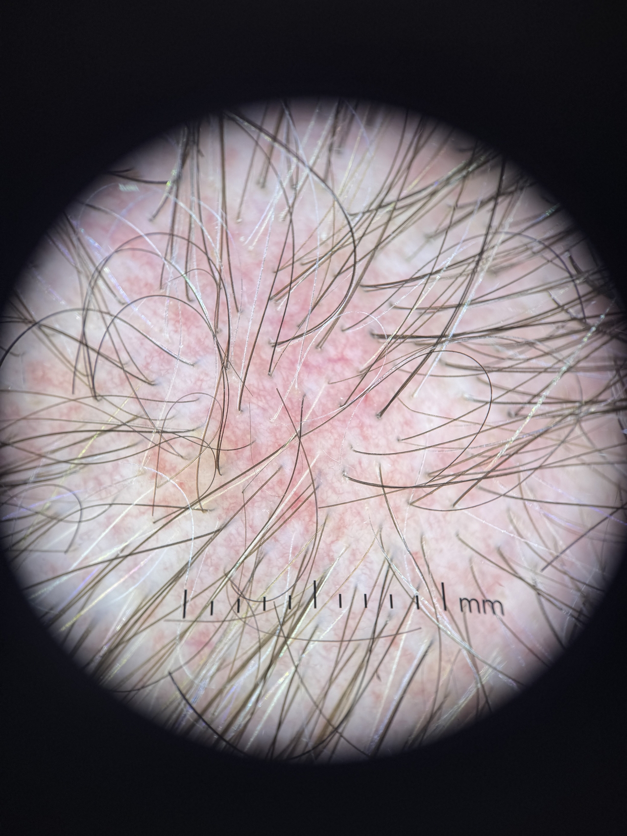
Microscopy
Between dermoscopy and reflectance confocal microscopy lies a middle ground of high-resolution, high magnification imaging acquired at video rate in a clinically-acceptable manner (short timeframe, lack of index-matching). We are exploring this space with the development of new low-cost microscopy tools to view blood vessel morphology for application to skin cancer detection, diagnosis of inflammatory skin diseases, and visualization of skin adnexae.
Devices
Devices for use in dermatology run the gamut from highly-engineered, low-cost tools which form the backbone of dermatologic practice (biopsy punches, cryotherapy spray cans, sutures) to high-cost, energy-based devices such as lasers and radiofrequency devices that are used primarily for cosmetic purposes. Most energy-based devices operate by selective destruction or ablation of a particular skin compartment (i.e. targeting particular adnexae or anatomic layers of the skin). We seek to find alternative methods for reproducing selective injury in particular compartments or geometries using low-cost, ubiquitous methods.