Research

The Kim Laboratory investigates molecular and physiological mechanisms that regulate glucose and energy homeostasis, with a focus on pathways that are disrupted in obesity, type 2 diabetes and fatty liver disease. We concentrate on key signaling nodes, including the kinase ROCK1, the lipoprotein receptor LRP1, and hepatokines such as clusterin (ApoJ), that link liver, adipose tissue, skeletal muscle and the brain. Our approaches integrate mouse genetics, physiology and omics.

1. Central regulation & leptin transport (brain–body axis)

We study how brain barrier sites and hypothalamic circuits sense circulating hormones and coordinate energy balance.

Leptin entry mechanism: Work in choroid-plexus epithelial cells has identified a complex between short-form leptin receptors (LepR) and LRP1 that mediates transport of circulating leptin into the cerebrospinal fluid. Disruption of this complex reduces leptin entry into the brain, diminishes hypothalamic STAT3 signaling and promotes hyperphagia and weight gain, providing a mechanism for central leptin resistance in obesity.

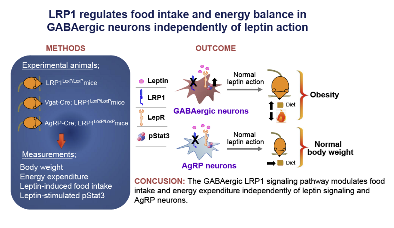
Hypothalamic signaling: Ongoing studies examine how hypothalamic Rho-kinase (ROCK1) and GABAergic circuits contribute to the regulation of food intake and energy expenditure.

2. ROCK1 in insulin resistance and fatty liver disease

We investigate ROCK1 as a stress-responsive kinase that links overnutrition to impaired insulin action and lipid accumulation.

Systemic insulin resistance: In liver, adipose tissue and pancreatic β-cells, ROCK1 activity interferes with insulin signaling and promotes de novo lipogenesis in the setting of high-fat feeding.

Therapeutic modulation: Genetic and pharmacologic studies in mouse models indicate that reducing ROCK1 activity improves insulin sensitivity and attenuates diet-induced fatty liver disease (NAFLD), supporting ROCK1 as a potential target in type 2 diabetes and related metabolic disorders.

3. Hepatokines and inter-organ crosstalk

We examine how liver-derived factors signal to peripheral tissues to shape whole-body metabolism.

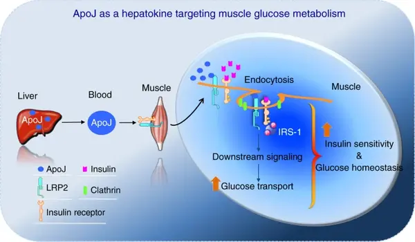
Liver–muscle axis: Work from the lab has identified clusterin (ApoJ) as a hepatokine that acts through the muscle receptor LRP2 to regulate insulin receptor internalization, insulin signaling and glucose uptake in skeletal muscle, linking altered ApoJ–LRP2 signaling to systemic insulin resistance.

Secretome analysis: Additional projects use genetic mouse models, secretome profiling and in vivo metabolic phenotyping to map liver–muscle and liver–brain communication pathways and to define inter-organ nodes that may be leveraged for metabolic therapy.

Brain & LRP1

GABA

Regulation of Energy Balance by Hypothalamic LRP1 This model illustrates our finding that LRP1 in GABAergic neurons is essential for regulating food intake and energy homeostasis. LRP1 deficiency in these neurons leads to metabolic obesity, highlighting a critical central pathway for body weight regulation.


Kang MC, Seo JA, Lee H, et al. LRP1 regulates food intake and energy balance in GABAergic neurons independently of leptin action. Am J Physiol Endocrinol Metab. 2021;320(2):E379-E389. doi:10.1152/ajpendo.00399.2020

ROCK1 & Insulin Secretion

s

ROCK1 regulates Insulin Secretion via Pyruvate Kinase This model illustrates a novel mechanism where ROCK1 acts as an upstream regulator of glycolysis in β-cells. Upon glucose stimulation, ROCK1 binds to Pyruvate Kinase (PK), facilitating ATP production and subsequent calcium influx, which are requisite for efficient insulin secretion.

Sung BJ, Lim SB, Yang WM, et al. ROCK1 regulates insulin secretion from β-cells. Mol Metab. 2022;66:101625. doi:10.1016/j.molmet.2022.101625

Hepatokines & Inter-organ Crosstalk

ApoJ

Apolipoprotein J is a Hepatokine Regulating Muscle Glucose Metabolism This schematic illustrates our discovery that Apolipoprotein J (ApoJ/Clusterin) acts as a hepatokine. It travels from the liver to skeletal muscle, regulating glucose metabolism and insulin sensitivity via the LRP2 receptor.


Seo JA, Kang MC, Yang WM, et al. Author Correction: Apolipoprotein J is a hepatokine regulating muscle glucose metabolism and insulin sensitivity. Nat Commun. 2020;11(1):2276. Published 2020 May 5. doi:10.1038/s41467-020-16305-6

Recent Publications

  • Lim, Soo, Sung Hee Choi, Hayley Shin, Bong Jun Cho, Ho Seon Park, Byung Yong Ahn, Seon Mee Kang, et al. (2012) 2012. “Effect of a Dipeptidyl Peptidase-IV Inhibitor, Des-Fluoro-Sitagliptin, on Neointimal Formation After Balloon Injury in Rats.”. PloS One 7 (4): e35007. https://doi.org/10.1371/journal.pone.0035007.

    BACKGROUND: Recently, it has been suggested that enhancement of incretin effect improves cardiac function. We investigated the effect of a DPP-IV inhibitor, des-fluoro-sitagliptin, in reducing occurrence of restenosis in carotid artery in response to balloon injury and the related mechanisms.

    METHODS AND FINDINGS: Otsuka Long-Evans Tokushima Fatty rats were grouped into four: control (normal saline) and sitagliptin 100, 250 and 500 mg/kg per day (n = 10 per group). Sitagliptin or normal saline were given orally from 1 week before to 2 weeks after carotid injury. After 3 weeks of treatment, sitagliptin treatment caused a significant and dose-dependent reduction in intima-media ratio (IMR) in obese diabetic rats. This effect was accompanied by improved glucose homeostasis, decreased circulating levels of high-sensitivity C-reactive protein (hsCRP) and increased adiponectin level. Moreover, decreased IMR was correlated significantly with reduced hsCRP, tumor necrosis factor-α and monocyte chemoattractant protein-1 levels and plasminogen activator inhibitor-1 activity. In vitro evidence with vascular smooth muscle cells (VSMCs) demonstrated that proliferation and migration were decreased significantly after sitagliptin treatment. In addition, sitagliptin increased caspase-3 activity and decreased monocyte adhesion and NFκB activation in VSMCs.

    CONCLUSIONS: Sitagliptin has protective properties against restenosis after carotid injury and therapeutic implications for treating macrovascular complications of diabetes.

  • Chu, Sang Hui, Ji-Hye Park, Mi Kyung Lee, Yoonsuk Jekal, Ki Yong Ahn, Jae Youn Chung, Dong Hoon Lee, et al. (2012) 2012. “The Association Between Pentraxin 3 and Insulin Resistance in Obese Children at Baseline and After Physical Activity Intervention.”. Clinica Chimica Acta; International Journal of Clinical Chemistry 413 (19-20): 1430-7. https://doi.org/10.1016/j.cca.2012.06.002.

    BACKGROUND: The role of pentraxin-3 (PTX3) in the development of insulin resistance is still not clear. We aimed to test 1) whether circulating PTX3 levels are associated with insulin resistance and 2) whether changes in PTX3 levels after the physical activity are associated with changes in insulin resistance.

    METHODS: Fifty-seven overweight or obese children (39 boys, 18 girls; age: 12.04±0.82y, BMI: 26.5±1.2 kg/m²) participated in the study. All participants were housed together and their amount of physical activity (1823.5±1.34 kcal/day) and food intake (1882±68.8 kcal/day) were tightly controlled.

    RESULTS: Circulating PTX3 levels at baseline were negatively associated with fasting insulin (r=-.336, p=0.012) and homeostasis model assessment of insulin resistance (HOMA-IR) (r=-.334, p=0.014) even after adjustment for BMI and Tanner stage. The degree of change in PTX3 levels notably associated with changes in fasting insulin (r=-.280, p=0.035) and HOMA-IR (r=-.281, p=.034) in response to the physical activity intervention. Subgroup analysis further indicates that HOMA-IR was improved more in subjects whose PTX3 levels were increased compared with subjects who PTX3 levels were decreased (HOMA-IR delta: -2.33±1.3 vs -1.46±0.70, p=0.004).

    CONCLUSION: PTX3 is negatively associated with insulin resistance and associated with changes in insulin resistance induced by physical activity in overweight and obese children.

  • Huang, Hu, Dong Kong, Kyung Hee Byun, Chianping Ye, Shuichi Koda, Dae Ho Lee, Byung-Chul Oh, et al. (2012) 2012. “Rho-Kinase Regulates Energy Balance by Targeting Hypothalamic Leptin Receptor Signaling.”. Nature Neuroscience 15 (10): 1391-8. https://doi.org/10.1038/nn.3207.

    Leptin regulates energy balance. However, knowledge of the critical intracellular transducers of leptin signaling remains incomplete. We found that Rho-kinase 1 (ROCK1) regulates leptin action on body weight homeostasis by activating JAK2, an initial trigger of leptin receptor signaling. Leptin promoted the physical interaction of JAK2 and ROCK1, thereby increasing phosphorylation of JAK2 and downstream activation of Stat3 and FOXO1. Mice lacking ROCK1 in either pro-opiomelanocortin (POMC) or agouti-related protein neurons, mediators of leptin action, displayed obesity and impaired leptin sensitivity. In addition, deletion of ROCK1 in the arcuate nucleus markedly enhanced food intake, resulting in severe obesity. Notably, ROCK1 was a specific mediator of leptin, but not insulin, regulation of POMC neuronal activity. Our data identify ROCK1 as a key regulator of leptin action on energy homeostasis.

  • Manabe, Yasuko, Katja S C Gollisch, Laura Holton, Young-Bum Kim, Josef Brandauer, Nobuharu L Fujii, Michael F Hirshman, and Laurie J Goodyear. (2013) 2013. “Exercise Training-Induced Adaptations Associated With Increases in Skeletal Muscle Glycogen Content.”. The FEBS Journal 280 (3): 916-26. https://doi.org/10.1111/febs.12085.

    Chronic exercise training results in numerous skeletal muscle adaptations, including increases in insulin sensitivity and glycogen content. To understand the mechanism leading to increased muscle glycogen, we studied the effects of exercise training on glycogen regulatory proteins in rat skeletal muscle. Female Sprague Dawley rats performed voluntary wheel running for 1, 4 or 7 weeks. After 7 weeks of training, insulin-stimulated glucose uptake was increased in epitrochlearis muscle. As compared with sedentary control rats, muscle glycogen did not change after 1 week of training, but increased significantly after 4 and 7 weeks. The increases in muscle glycogen were accompanied by elevated glycogen synthase activity and protein expression. To assess the regulation of glycogen synthase, we examined its major activator, protein phosphatase 1 (PP1), and its major deactivator, glycogen synthase kinase (GSK)-3. Consistent with glycogen synthase activity, PP1 activity was unchanged after 1 week of training but significantly increased after 4 and 7 weeks of training. Protein expression of R(GL)(G(M)), another regulatory PP1 subunit, significantly decreased after 4 and 7 weeks of training. Unlike PP1 activity, GSK-3 phosphorylation did not follow the pattern of glycogen synthase activity. The   40% decrease in GSK-3α phosphorylation after 1 week of exercise training persisted until 7 weeks, and may function as a negative feedback mechanism in response to elevated glycogen. Our findings suggest that exercise training-induced increases in muscle glycogen content could be regulated by multiple mechanisms, including enhanced insulin sensitivity, glycogen synthase expression, allosteric activation of glycogen synthase, and PP1 activity.

  • Kim, Young Bum, Ji Hye Hwang, Tae Won Kim, Hyun Ju Chang, and Sang Gil Lee. (2012) 2012. “Would Complex Decongestive Therapy Reveal Long Term Effect and Lymphoscintigraphy Predict the Outcome of Lower-Limb Lymphedema Related to Gynecologic Cancer Treatment?”. Gynecologic Oncology 127 (3): 638-42. https://doi.org/10.1016/j.ygyno.2012.09.015.

    OBJECTIVE: The aims of this study were to investigate the long-term effect of complex decongestive therapy (CDT) on lymphedema volume reduction, especially considering the proximal and distal parts of the leg, and to evaluate the utility of pre-therapy lymphoscintigraphy in predicting the response to CDT in patients with lower-limb lymphedema after surgery for gynecologic cancer.

    METHODS: Medical records of 158 patients with secondary lymphedema of unilateral leg after surgery for gynecological cancer were reviewed retrospectively. They were treated with two weeks of CDT along with self-administered home therapy and were followed up for 24 months. Whole, proximal and distal leg volume was serially measured by using an optoelectric volumeter prior to and immediately after therapy, and follow-up visits at months 3, 6, 12 and 24. Lymphoscintigraphy was performed prior to therapy.

    RESULTS: The percent volume reduction was 22.1% in the whole leg, 30.9% in the distal leg and 18.4% in the proximal leg immediately after CDT. The volume reduction was maintained for 24 months, but the distal leg was significantly well maintained better than the proximal leg. Extremity radioisotope uptake ratio (EUR) among lymphoscintigraphic findings could predict the improvement of lymphedema volume in the distal, proximal and whole leg.

    CONCLUSIONS: This study suggests that the long-term edema reducing effects of CDT are better maintained in the distal leg than in the proximal part, and initial lymphoscintigraphic quantitative finding may usefully predict the short and long-term response to CDT.