Full PubMed Bibliography: https://pubmed.ncbi.nlm.nih.gov/?term=saper+cb&sort=date
Research
Wake-Sleep Circuitry
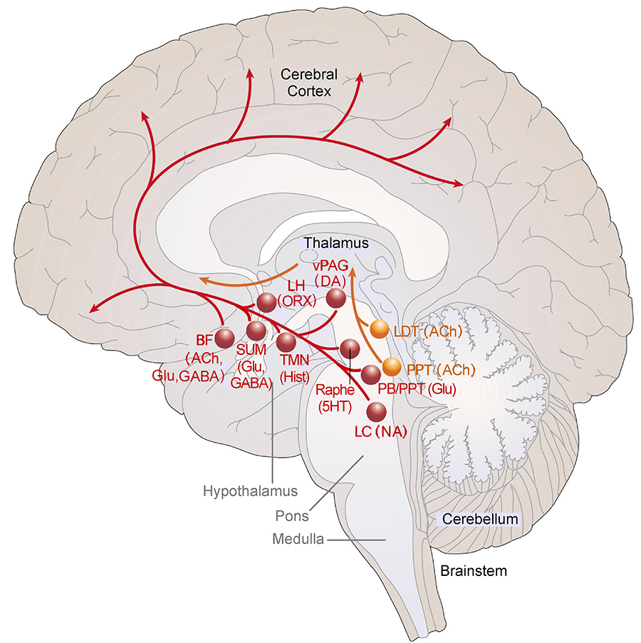
 A schematic diagram of the ascending arousal system.
We use cutting-edge methods including opto- and chemogenetics and calcium imaging in transgenic animals to identify the brain circuits that control wake-sleep states. These studies use single nucleus RNA sequencing to determine the genetically specified cell types that contribute to wake-sleep regulation, and to determine their neurotransmitters, targets, and receptors. We also are interested in the circuitry that insures breathing during sleep, and how manipulations of this circuitry may improve disorders such as sleep apnea and opioid-induced respiratory depression.
Brain Circuitry for Circadian Rhythms
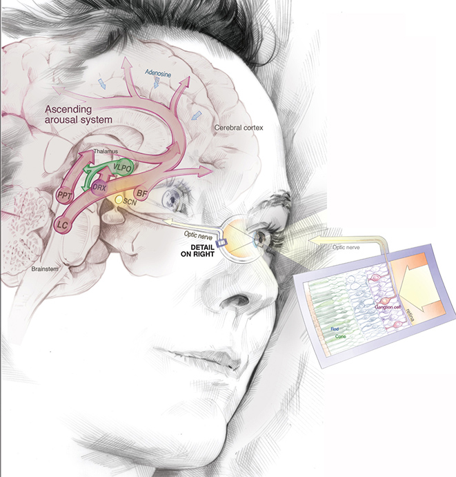
 A schematic diagram showing the pathway for light resetting the suprachiasmatic nucleus, and its outputs to the wake-sleep regulatory system.
We use the same approaches to identify circuitry used by the suprachiasmatic nucleus, the brain's biological clock, to control the daily rhythms of physiology and behavior, including wake-sleep cycles, body temperature, feeding, corticosteroid secretion, and behaviors such as aggression and sexual behavior.
Brain Circuitry for Thermoregulation
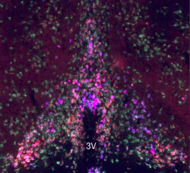
 Multi-plex in situ hybridization for genes marking preoptic neurons that contribute to bouts of torpor in mice, where they reduce body temperature and metabolism to survive in a cold environment when there is lack of food.
Body temperature is closely integrated with wake-sleep cycles, circadian rhythms, and a variety of behaviors such as feeding, stress, and reproduction. We use the same advanced methods as above to identify the genetically specified cell types that regulate body temperature, as well as their connections and neurotransmitters. We are particularly interested in the brain mechanisms for fever during inflammatory states and for torpor response in mice, which when placed in a cool environment with not enough food to eat, have a steep fall in body temperature, similar to hibernation.
Circuitry in Human Brains and Effects of Neurodegenerative Disorders
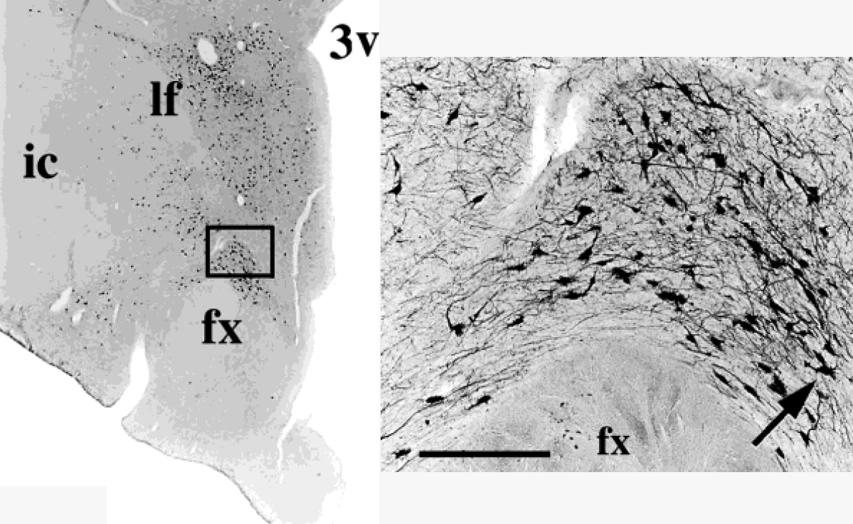
 Neurons in a human brain stained immunohistochemically for melanin-concentrating hormone
Many of the circuits we study in animals also contain the same neurotransmitters in humans. We use immunohistochemical staining to study these neurons in human post-mortem brains, and to determine how these neurons are affected by Alzheimer's, Parkinson's and other neurodegenerative diseases.
Laboratory Collaborations
Saper Lab current collaborators include:
Thomas Scammell Lab
Brad Lowell Lab
Mark Andermann Lab
Elda Arrigoni Lab
Veronique van der Horst Lab
Vetrivelan Ramalingam Lab
Satvinder Kaur Lab
Natalia Machado Lab
Roberto De Luca Lab
