Research

Liver Organoids

Liver Organoids

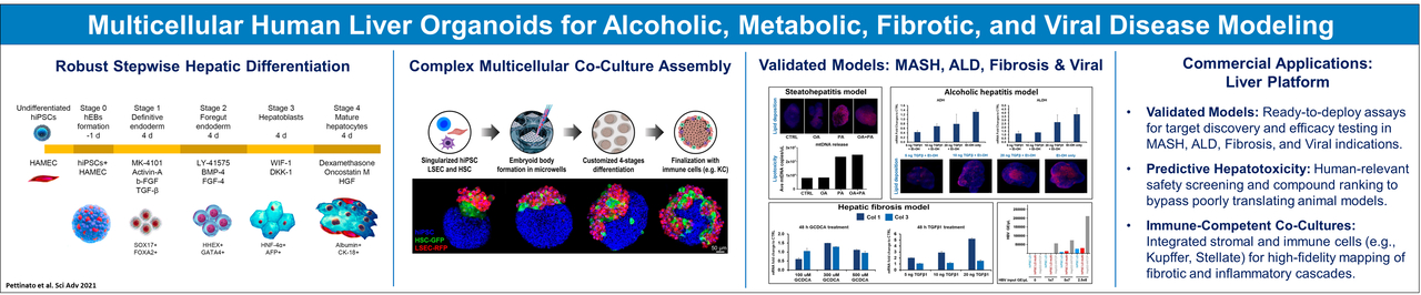
Liver organoids are three-dimensional, miniaturized liver-like structures grown from stem cells or primary liver cells in vitro. They mimic key aspects of liver function, including metabolism, detoxification, and albumin production. These organoids are used for disease modeling, drug testing, regenerative medicine, and studying liver development. In tissue engineering, liver organoids play a crucial role in developing bioengineered liver tissues for transplantation, offering a potential solution for liver disease treatment and organ shortages. Their ability to self-organize and replicate liver tissue architecture makes them a promising tool for biomedical research and regenerative medicine.

Pancreatic Organoids

Pancreatic Organoids

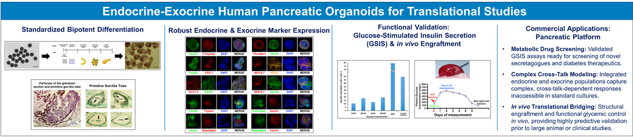
Pancreatic organoids are three-dimensional, miniaturized pancreatic tissue models grown from stem cells or primary pancreatic cells in vitro. They replicate key functions of the pancreas, including enzyme secretion and hormone production, making them valuable for studying pancreatic development, disease modeling (such as diabetes and pancreatic cancer), and drug testing. In tissue engineering, pancreatic organoids hold potential for regenerative therapies, offering new avenues for treating pancreatic disorders and developing bioengineered pancreatic tissues for transplantation.

Intestinal Organoids

Intestinal Organoids

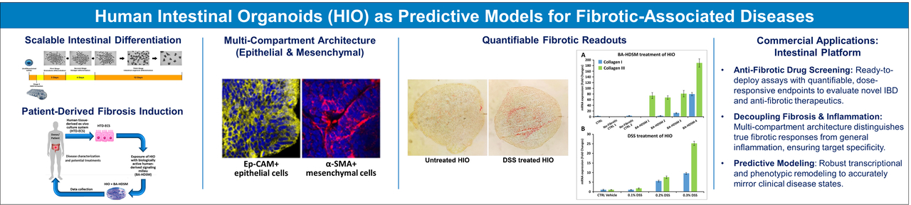
Intestinal organoids are miniature, lab-grown models of the intestine that closely resemble its structure and function. Developed from stem cells, they contain key cell types found in the gut and can perform essential processes like absorbing nutrients and maintaining a protective barrier. These organoids serve as powerful tools for studying diseases, testing drugs, and exploring interactions with the microbiome. In the field of tissue engineering, they offer promising possibilities for regenerating damaged intestinal tissue and developing new treatments for digestive disorders.

Recent Publications

  • Burrello, Nunziatina, Mario Salmeri, Anna Perdichizzi, Salvatore Bellanca, Giuseppe Pettinato, Rosario D’Agata, Enzo Vicari, and Aldo E Calogero. (2009) 2009. “Candida Albicans Experimental Infection: Effects on Human Sperm Motility, Mitochondrial Membrane Potential and Apoptosis.”. Reproductive Biomedicine Online 18 (4): 496-501.

    Studies suggest Candida albicans infection has a negative effect on sperm function, including fertilizing ability. Assisted reproduction treatment using spermatozoa from a patient with unrecognized C. albicans infection did not result in fertilization. Preliminary evidence suggested an effect on sperm motility and apoptosis. This study was undertaken to evaluate the effects of experimentally induced C. albicans infection on motility, membrane mitochondrial potential (MMP), chromatin packaging and apoptosis [membrane phosphatidylserine (PS) externalization and DNA fragmentation] of spermatozoa isolated from normozoospermic healthy men. Motile spermatozoa were isolated by swim-up from 13 normal volunteers and exposed to increasing concentrations (0, 1000, 10,000, and 100,000 cfu/ml) of the fungus for 3 and 24 h. C. albicans was isolated from vaginal swabs, after identification, freshly prepared for experiments. Following incubation, sperm motility decreased significantly (P < 0.05 from 10,000 cfu/ml) and spermatozoa with reduced MMP or PS externalization, an early sign of apoptosis, increased in a time- and concentration-dependent manner. Sperm DNA fragmentation and chromatin integrity increased slightly after exposure to C. albicans, but the increase did not reach statistical significance. This study showed that C. albicans infection may decrease the functional competence of spermatozoa by reducing motility and MMP and by promoting molecular apoptosis mechanisms.

  • Pettinato, Giuseppe, Wendy S Vanden Berg-Foels, Ning Zhang, and Xuejun Wen. (2014) 2014. “ROCK Inhibitor Is Not Required for Embryoid Body Formation from Singularized Human Embryonic Stem Cells.”. PloS One 9 (11): e100742. https://doi.org/10.1371/journal.pone.0100742.

    We report a technology to form human embryoid bodies (hEBs) from singularized human embryonic stem cells (hESCs) without the use of the p160 rho-associated coiled-coil kinase inhibitor (ROCKi) or centrifugation (spin). hEB formation was tested under four conditions: +ROCKi/+spin, +ROCKi/-spin, -ROCKi/+spin, and -ROCKi/-spin. Cell suspensions of BG01V/hOG and H9 hESC lines were pipetted into non-adherent hydrogel substrates containing defined microwell arrays. hEBs of consistent size and spherical geometry can be formed in each of the four conditions, including the -ROCKi/-spin condition. The hEBs formed under the -ROCKi/-spin condition differentiated to develop the three embryonic germ layers and tissues derived from each of the germ layers. This simplified hEB production technique offers homogeneity in hEB size and shape to support synchronous differentiation, elimination of the ROCKi xeno-factor and rate-limiting centrifugation treatment, and low-cost scalability, which will directly support automated, large-scale production of hEBs and hESC-derived cells needed for clinical, research, or therapeutic applications.

  • Pettinato, Giuseppe, Xuejun Wen, and Ning Zhang. (2014) 2014. “Formation of Well-Defined Embryoid Bodies from Dissociated Human Induced Pluripotent Stem Cells Using Microfabricated Cell-Repellent Microwell Arrays.”. Scientific Reports 4: 7402. https://doi.org/10.1038/srep07402.

    A simple, scalable, and reproducible technology that allows direct formation of large numbers of homogeneous and synchronized embryoid bodies (EBs) of defined sizes from dissociated human induced pluripotent stem cells (hiPSCs) was developed. Non-cell-adhesive hydrogels were used to create round-bottom microwells to host dissociated hiPSCs. No Rho-associated kinase inhibitor (ROCK-i), or centrifugation was needed and the side effects of ROCK-i can be avoided. The key requirement for the successful EB formation in addition to the non-cell-adhesive round-bottom microwells is the input cell density per microwell. Too few or too many cells loaded into the microwells will compromise the EB formation process. In parallel, we have tested our microwell-based system for homogeneous hEB formation from dissociated human embryonic stem cells (hESCs). Successful production of homogeneous hEBs from dissociated hESCs in the absence of ROCK-i and centrifugation was achieved within an optimal range of input cell density per microwell. Both the hiPSC- and hESC-derived hEBs expressed key proteins characteristic of all the three developmental germ layers, confirming their EB identity. This novel EB production technology may represent a versatile platform for the production of homogeneous EBs from dissociated human pluripotent stem cells (hPSCs).

  • Ramanathan, Rajesh, Giuseppe Pettinato, John T Beeston, David D Lee, Xuejun Wen, Martin J Mangino, and Robert A Fisher. (2015) 2015. “Transplantation of Human Stem Cell-Derived Hepatocytes in an Animal Model of Acute Liver Failure.”. Surgery 158 (2): 349-59. https://doi.org/10.1016/j.surg.2015.04.014.

    INTRODUCTION: Hepatocyte cell transplantation can be life-saving in patients with acute liver failure (ALF); however, primary human hepatocyte transplantation is limited by the scarcity of donor hepatocytes. We investigated the effect of stem cell-derived, hepatocyte-like cells in an animal xenotransplant model of ALF.

    METHODS: Intraperitoneal d-galactosamine was used to develop a lethal model of ALF in the rat. Human induced pluripotent stem cells (iPSC), human mesenchymal stem cells, and human iPSC combined with human endothelial cells (iPSC + EC) were differentiated into hepatocyte-like cells and transplanted into the spleens of athymic nude rats with ALF.

    RESULTS: A reproducible lethal model of ALF was achieved with nearly 90% death within 3 days. Compared with negative controls, rats transplanted with stem cell-derived, hepatocyte-like cells were associated with increased survival. Human albumin was detected in the rat serum 3 days after transplantation in more than one-half the animals transplanted with hepatocyte-like cells. Only animals transplanted with iPSC + EC-derived hepatocytes had serum human albumin at 14 days posttransplant. Transplanted hepatocyte-like cells homed to the injured rat liver, whereas the ECs were only detected in the spleen.

    CONCLUSION: Transplantation of stem cell-derived, hepatocyte-like cells improved survival with evidence of in vivo human albumin production. Combining ECs may prolong cell function after transplantation.

  • Pettinato, Giuseppe, Rajesh Ramanathan, Robert A Fisher, Martin J Mangino, Ning Zhang, and Xuejun Wen. (2016) 2016. “Scalable Differentiation of Human IPSCs in a Multicellular Spheroid-Based 3D Culture into Hepatocyte-Like Cells through Direct Wnt/β-Catenin Pathway Inhibition.”. Scientific Reports 6: 32888. https://doi.org/10.1038/srep32888.

    Treatment of acute liver failure by cell transplantation is hindered by a shortage of human hepatocytes. Current protocols for hepatic differentiation of human induced pluripotent stem cells (hiPSCs) result in low yields, cellular heterogeneity, and limited scalability. In the present study, we have developed a novel multicellular spheroid-based hepatic differentiation protocol starting from embryoid bodies of hiPSCs (hiPSC-EBs) for robust mass production of human hepatocyte-like cells (HLCs) using two novel inhibitors of the Wnt pathway. The resultant hiPSC-EB-HLCs expressed liver-specific genes, secreted hepatic proteins such as Albumin, Alpha Fetoprotein, and Fibrinogen, metabolized ammonia, and displayed cytochrome P450 activities and functional activities typical of mature primary hepatocytes, such as LDL storage and uptake, ICG uptake and release, and glycogen storage. Cell transplantation of hiPSC-EB-HLC in a rat model of acute liver failure significantly prolonged the mean survival time and resolved the liver injury when compared to the no-transplantation control animals. The transplanted hiPSC-EB-HLCs secreted human albumin into the host plasma throughout the examination period (2 weeks). Transplantation successfully bridged the animals through the critical period for survival after acute liver failure, providing promising clues of integration and full in vivo functionality of these cells after treatment with WIF-1 and DKK-1.

  • Pettinato, Giuseppe, Xuejun Wen, and Ning Zhang. (2015) 2015. “Engineering Strategies for the Formation of Embryoid Bodies from Human Pluripotent Stem Cells.”. Stem Cells and Development 24 (14): 1595-609. https://doi.org/10.1089/scd.2014.0427.

    Human pluripotent stem cells (hPSCs) are powerful tools for regenerative therapy and studying human developmental biology, attributing to their ability to differentiate into many functional cell types in the body. The main challenge in realizing hPSC potential is to guide their differentiation in a well-controlled manner. One way to control the cell differentiation process is to recapitulate during in vitro culture the key events in embryogenesis to obtain the three developmental germ layers from which all cell types arise. To achieve this goal, many techniques have been tested to obtain a cellular cluster, an embryoid body (EB), from both mouse and hPSCs. Generation of EBs that are homogeneous in size and shape would allow directed hPSC differentiation into desired cell types in a more synchronous manner and define the roles of cell-cell interaction and spatial organization in lineage specification in a setting similar to in vivo embryonic development. However, previous success in uniform EB formation from mouse PSCs cannot be extrapolated to hPSCs possibly due to the destabilization of adherens junctions on cell surfaces during the dissociation into single cells, making hPSCs extremely vulnerable to cell death. Recently, new advances have emerged to form uniform human embryoid bodies (hEBs) from dissociated single cells of hPSCs. In this review, the existing methods for hEB production from hPSCs and the results on the downstream differentiation of the hEBs are described with emphases on the efficiency, homogeneity, scalability, and reproducibility of the hEB formation process and the yield in terminal differentiation. New trends in hEB production and directed differentiation are discussed.

SCORE Center
Beth Israel Deaconess Medical Center
Research North, Room RN0245
99 Brookline Avenue
Boston MA, 02215