The Preclinical MRI Core at Beth Israel Deaconess Medical Center offers instrumentation and expertise for a broad range of magnetic resonance imaging and spectroscopy applications for small animals and tissue samples.
The Preclinical MRI Core is a full-service facility that provides protocol design, image acquisition, and data analysis.
About Our Standard Lab Applications
The lab maintains a state-of-the-art small animal MRI scanner operating at 9.4 Tesla field strength (Bruker Biospec 94/20) in addition to a DNP carbon-13 hyperpolarizer.
We run imaging and spectroscopy experiments on an hourly fee basis for BIDMC colleagues and outside investigators. We will be happy to discuss how MR imaging can be used to further your research goals.
Standard applications of the lab include anatomic, T1, T2, diffusion, fMRI, perfusion, metabolism, and ex-vivo tissue imaging.
-
In Vivo Imaging: Anatomic in vivo imaging of the head, thorax, and abdomen.
-
Patient Tissue Samples: Postsurgical tissue samples can be imaged at very high resolution using our high-field MRI.
-
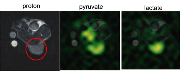
Hyperpolarized 13C: Tissue metabolism and perfusion can be imaged using hyperpolarized carbon-13 tracers.
-
Cardiac Imaging: Ejection fraction, ventricle wall motion, and strain can be imaged and quantified.
-
Blood Flow: Quantitative imaging of tissue perfusion with arterial spin-labeling or hyperpolarized carbon-13.
-
Angiography: Angiographic images can also be obtained with flow-compensated gradient echo imaging.
-
Image Analysis: Images can be analyzed to provide quantitative maps of diagnostic physiological parameters, including blood flow and metabolism.
Preclinical MRI Core Pricing
Request a Scheduling Slot
Please email Aaron Grant, PhD, with the following information:
- Date needed
- Start and end times
- Desired protocol
- # samples/animals
Core Director
Aaron K. Grant, PhD
P: 617-667-3265
F: 617-667-7917
Assistant Director
Gopal Varma, PhD
P: 617-667-0281