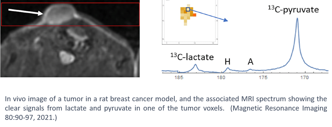
Hyperpolarization is a technology for aligning, or polarizing, the nuclear spins in a contrast agent or metabolic tracer. Agents prepared this way can dramatically enhance nuclear magnetization and, as a result, produce MR signals up to 100,000 times stronger versus conventional imaging. This makes it possible to image the transport, uptake, and metabolic transformations of tracer molecules non-invasively and in real-time. The large signal gains offered by hyperpolarization make it possible to image tissue function and metabolism in fundamentally new ways.
Using this technology, we have investigated several aspects of tumor metabolism that may be critical in therapeutic discovery and research.
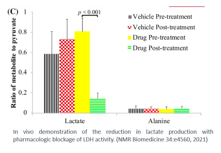
One approach to tumor therapeutics is to cut off the tumor metabolic processes that sustain tumor growth. Tumors utilize large quantities of glucose as a metabolic source, with one of the end steps being pyruvate-to-lactate conversion. Therefore, the elimination of lactate dehydrogenase (LDH), the enzyme needed for this end step, is one therapeutic option.

In animal tumor models, we demonstrated that pharmacologically blocking LDH resulted in a large drop in the lactate-to-pyruvate ratio (see graph to the right), demonstrating that the completion of the metabolic pathway was effectively inhibited and could be monitored in vivo. This demonstrates the potential opportunity to study this important therapeutic pathway under clinical conditions.
Ablation therapy, involving the destruction of tumors through direct application of localized heat, is an option for tumors that are not surgical candidates. Unfortunately, ablation sometimes results in increased growth of distal tumors. In a rat tumor model, hyperpolarized MRI was able to show the increased lactate production in remote tumors with ablation of a primary tumor, while treatment with an inhibitor to the cell surface receptor c-MET eliminated this increase of lactate.

In a mouse model of gastric cancer, hyperpolarized MRI was able to detect response to treatment within 4 days of initiation of treatment, and before the response was observable on standard MRI or PET imaging (Molec Imaging Biol, 2022).
Together, these studies demonstrate the potential of hyperpolarized MRI to detect tumor response to therapeutic interventions in vivo, which may be used for non-invasive monitoring and optimization of treatment.
