Autonomic Control of Kidney Function

Prior work by others suggests that renal nerves arise from several neuron subpopulations in sympathetic ganglia. It is possible that these specific subpopulations of neurons, as well as the CNS circuits that innervate them selectively control tubular transport, glomerular filtration rate or renin release. This NIH funded project aims to create the tools to define the individual subpopulations of brainstem neurons which connect to the preganglionic neurons that control sympathetic postganglionic neurons important for renal function.

Neurons originating from the paraventricular hypothalamus (PVH) and other hypothalamic nuclei, the medullary raphe pallidus, as well as the rostral ventrolateral medulla (RVLM) and the A5 cell group in the pons project to the intermediolateral column (IML) at the T8 – T12 spinal cord, where nearly all renal sympathetic preganglionic fibers originate.

Combined, PVH, RVLM and A5 neurons provide the vast majority of brain input to the spinal cord, and the proposed studies will focus on neuron subpopulations in these brain areas which control kidney function.
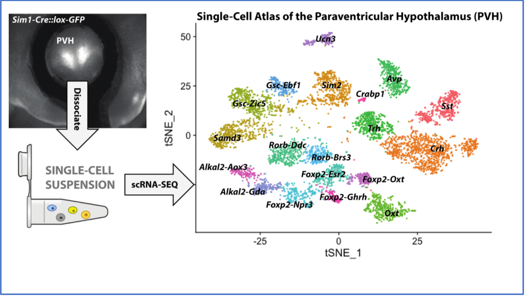


single nuclei RNA sequencing
Single-nucleus RNA sequencing of paraventricular hypothalamus (PVH) neurons. A reporter mouse expressing green fluorescence protein just in cells of the PVH (top left), had the fluorescent region recovered. The isolated PVH was then dissociated into single cell suspensions, cells separated by microfluidic device, and then each individual cell or nucleus was sequenced and analyzed. Neuron subpopulations are visualized using UMAP projection and expression of marker genes that define each cluster are indicated in the tSNE plot. Each dot represents a single sequenced nucleus.

 
laser stimulation of light sensitive cells in brain causes blood pressure rise
Optogenetic stimulation of RVLM leads to increased renal nerve activity and blood pressure.  Schematic on the left shows optogenetic stimulation of C1 neurons of RVLM and monitoring of renal activity and blood pressure. Channel rhodopsin (ChR2) was expressed in RVLM of DBH-cre mouse. Optogenetic stimulation of these neurons with blue laser increases the renal nerve activity and blood pressure, which reverts to baseline when stimulation is stopped (right panel).