Micro-computed tomography (microCT) is an imaging technique that uses x-rays to produce cross-sectional images of an object that can be reconstructed to create a three-dimensional model. It allows for non-destructive quantitative analysis of the density, geometry and microarchitecture of mineralized or high-density material, particularly bone and biomaterials stained with contrast chemicals. The reason the technique is called micro-computed tomography is that the pixels are in the micrometer range; much smaller than conventional clinical computed tomography (CT) scanners. However, with an increase in resolution comes a decrease in the field of view that can be imaged. This means that microCT can only be performed on small specimens such as human biopsies or animal bones.
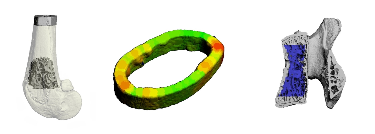
Our lab offers ex vivo microCT imaging services for the assessment of bone microarchitecture and mineral density. Our facility has two Scanco µCT40 scanners that allow for scanning at resolutions down to 6 μm3 and can scan specimens up to 36 mm in diameter and 75 mm in length. Our expert staff work closely with academic and industry clients to meet their skeletal phenotyping needs. Typical analyses that we perform include measuring the trabecular microarchitecture and cortical morphology of bones, however we can customize our analysis to meet a client’s needs. Bones that we typically analyze in rodents and other small animals include the femur, tibia, and lumbar vertebrae, however other regions can also be analyzed based on the focus area of the study. At the completion of our analysis, we provide clients with the microCT results organized in a spreadsheet, 2D and 3D images of samples, and a short report summarizing the results and methods for the study. We also provide clients with consultation on the interpretation of the microCT results. If you are interested in learning more about using microCT for the assessment of bones, you can find more information in the guidelines that were written by Dr. Bouxsein and colleagues